

- Featured in Physics
- Open Access
Second-Order Microscopic Nonlinear Optical Susceptibility in a Centrosymmetric Material: Application to Imaging Valence Electron Motion
Phys. Rev. X 16, 011006 – Published 7 January, 2026
DOI: https://doi.org/10.1103/7vqw-jbs7
Abstract
We report measurements of phase-matched nonlinear x-ray and optical mixing from single-crystal silicon using subresonant 0.95 eV laser pulses and 9.5 keV hard x-ray pulses from the Linear Coherent Light Source free-electron laser. The mixing signal appears as energy and momentum sidebands to the elastic Bragg peak. It is proportional to the magnitude squared of the relevant temporal and spatial Fourier components of the optically induced microscopic charges and currents. We measure the first- and second-order sideband to the 220 Bragg peak and find that the efficiency is maximized when the applied field is along the reciprocal lattice vector. For an optical intensity of approximately , we measure peak efficiencies of and for the first- and second-order sideband, respectively (relative to the elastic Bragg peak). The first-order sideband is consistent with induced microscopic currents along the applied electric field and an isotropic response. The second-order sideband depends nontrivially on the optical field orientation and is consistent with an anisotropic response originating from induced charges along the bonds with site symmetry. The results agree well with first-principles Floquet-Bloch calculations.
Physics Subject Headings (PhySH)
 synopsis

X-Ray–Optical Technique Detects Subtle Signal
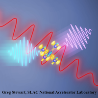
A high-sensitivity measurement reveals the asymmetric shifts of electrons along atomic bonds.
See more in Physics
Popular Summary
The detailed valence-electron density of a material plays a central role in its structure and properties. Measuring its atomic-scale structure is challenging because the valence density tends to be delocalized and is a small fraction of the total electron density. We isolated the optically driven valence-electron density in silicon using a nonlinear version of x-ray diffraction. From a single nonlinear Bragg peak corresponding to oscillations at twice the optical frequency, we isolated the valence density along the tetrahedral bonds without requiring phase information. While this nonlinear optical response is forbidden macroscopically, the angstrom-scale resolution provided by a hard x-ray probe allows sensitivity to atomic-scale inversion symmetry breaking, and thus local nonlinear optical responses that are invisible to optical measurements. The measurements agree well with first-principles calculations. The results have implications for studying nontrivial electronic ordering in more complex materials, both near and far from equilibrium.
Article Text
References (34)
- N. W. Ashcroft and N. D. Mermin, Solid State Physics (Holt-Saunders, New York, 1976).
- R. M. Martin, Lattice vibrations in silicon: Microscopic dielectric model, Phys. Rev. Lett. 21, 536 (1968).
- R. M. Martin, Dielectric screening model for lattice vibrations of diamond-structure crystals, Phys. Rev. 186, 871 (1969).
- R. W. James, The Optical Principles of the Diffraction of X-rays (G. Bell, London, 1948) (original edition).
- J. B. Roberto, B. W. Batterman, and D. T. Keating, Diffraction studies of the (222) reflection in Ge and Si: Anharmonicity and the bonding electron, Phys. Rev. B 9, 2590 (1974).
- J. Hastings and B. Batterman, High-order anharmonic forbidden neutron reflections in silicon, Phys. Rev. B 12, 5580 (1975).
- Y. Yang and P. Coppens, On the experimental electron distribution in silicon, Solid State Commun. 15, 1555 (1974).
- Z. W. Lu, A. Zunger, and M. Deutsch, Electronic charge distribution in crystalline diamond, silicon, and germanium, Phys. Rev. B 47, 9385 (1993).
- I. Freund and B. Levine, Optically modulated x-ray diffraction, Phys. Rev. Lett. 25, 1241 (1970).
- P. Eisenberger and S. McCall, Mixing of x-ray and optical photons, Phys. Rev. A 3, 1145 (1971).
- T. E. Glover, D. Fritz, M. Cammarata, T. Allison, S. Coh, J. Feldkamp, H. Lemke, D. Zhu, Y. Feng, R. Coffee et al., X-ray and optical wave mixing, Nature (London) 488, 603 (2012).
- D. Popova-Gorelova, D. A. Reis, and R. Santra, Theory of x-ray scattering from laser-driven electronic systems, Phys. Rev. B 98, 224302 (2018).
- D. Popova-Gorelova and R. Santra, Atomic-scale imaging of laser-driven electron dynamics in solids, Commun. Phys. 7, 317 (2024).
- C. Boemer, D. Krebs, A. Benediktovitch, E. Rossi, S. Huotari, and N. Rohringer, Towards novel probes for valence charges via x-ray optical wave mixing, Faraday Discuss. 228, 451 (2021).
- D. Krebs and N. Rohringer, Theory of parametric x-ray optical wavemixing processes, arXiv:2104.05838.
- K. Tamasaku, K. Sawada, E. Nishibori, and T. Ishikawa, Visualizing the local optical response to extreme-ultraviolet radiation with a resolution of , Nat. Phys. 7, 705 (2011).
- A. Schori, C. Bömer, D. Borodin, S. P. Collins, B. Detlefs, M. Moretti Sala, S. Yudovich, and S. Shwartz, Parametric down-conversion of x rays into the optical regime, Phys. Rev. Lett. 119, 253902 (2017).
- S. Sofer, O. Sefi, E. Strizhevsky, H. Aknin, S. P. Collins, G. Nisbet, B. Detlefs, C. J. Sahle, and S. Shwartz, Observation of strong nonlinear interactions in parametric down-conversion of x-rays into ultraviolet radiation, Nat. Commun. 10, 5673 (2019).
- J. D. Jackson, Classical Electrodynamics (John Wiley & Sons, New York, 1998).
- M. Chollet, R. Alonso-Mori, M. Cammarata, D. Damiani, J. Defever, J. T. Delor, Y. Feng, J. M. Glownia, J. B. Langton, S. Nelson et al., The x-ray pump-probe instrument at the linac coherent light source, Synchrotron Radiat. News 22, 503 (2015).
- N. Hartmann, W. Helml, A. Galler, M. R. Bionta, J. Grünert, S. L. Molodtsov, K. Ferguson, S. Schorb, M. Swiggers, S. Carron et al., Sub-femtosecond precision measurement of relative x-ray arrival time for free-electron lasers, Nat. Photonics 8, 706 (2014).
- K. Momma and F. Izumi, vesta3 for three-dimensional visualization of crystal, volumetric and morphology data, J. Appl. Crystallogr. 44, 1272 (2011).
- G. Bricogne, Maximum entropy and the foundations of direct methods, Found. Crystallogr. 40, 410 (1984).
- E. Weckert and K. Hümmer, Multiple-beam x-ray diffraction for physical determination of reflection phases and its applications, Found. Crystallogr. 53, 108 (1997).
- S. Ghimire and D. A. Reis, High-harmonic generation from solids, Nat. Phys. 15, 10 (2019).
- D. Basov, R. Averitt, and D. Hsieh, Towards properties on demand in quantum materials, Nat. Mater. 16, 1077 (2017).
- S. Huang, Y. Ding, Y. Feng, E. Hemsing, Z. Huang, J. Krzywinski, A. A. Lutman, A. Marinelli, T. J. Maxwell, and D. Zhu, Generating single-spike hard x-ray pulses with nonlinear bunch compression in free-electron lasers, Phys. Rev. Lett. 119, 154801 (2017).
- J. Yan, W. Qin, Y. Chen, W. Decking, P. Dijkstal, M. Guetg, I. Inoue, N. Kujala, S. Liu, T. Long, N. Mirian, and G. Geloni, Terawatt-attosecond hard x-ray free-electron laser at high repetition rate, Nat. Photonics 18, 1293 (2024).
- D. Zhu and D. A. Reis, Attosecond x-ray laser vision, Nat. Photonics 18, 1232 (2024).
- D. Popova-Gorelova and R. Santra, Microscopic nonlinear optical response: Analysis and calculations with the Floquet-Bloch formalism, Struct. Dyn. 11, 014102 (2024).
- Z. H. Levine and D. C. Allan, Linear optical response in silicon and germanium including self-energy effects, Phys. Rev. Lett. 63, 1719 (1989).
- J. Phillips, Band structure of silicon, germanium, and related semiconductors, Phys. Rev. 125, 1931 (1962).
- M. S. Hybertsen and S. G. Louie, First-principles theory of quasiparticles: Calculation of band gaps in semiconductors and insulators, Phys. Rev. Lett. 55, 1418 (1985).
- B. E. Warren, X-Ray Diffraction (Courier Corporation, New York, 1990).

