

- Open Access
Cell Bulging and Extrusion in a Three-Dimensional Bubbly Vertex Model for Curved Epithelial Sheets
Phys. Rev. X 16, 021023 – Published 30 April, 2026
DOI: https://doi.org/10.1103/x82g-cq7n
Abstract
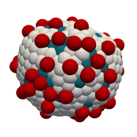
Cell extrusion is an essential mechanism for controlling cell density in epithelial tissues. Another essential element of epithelia is curvature, which is required to achieve complex shapes, like in the lung or intestine. Here, we introduce a three-dimensional bubbly vertex model to study the interplay between extrusion and curvature. We find a generic cellular bulging instability at topological defects, which is much stronger than for standard vertex models. Analyzing cell shapes in three-dimensional imaging data of spherical mouse colon organoids, we infer that pentagonal cells have an increased basal interfacial tension, suggesting that cells at topological defects react to the different force conditions. Using the bubbly vertex model, we show that such basal tensions stabilize against the predicted instability and result in better cell shape control than tissue-scale mechanisms such as lumen pressure and spontaneous curvature. Our theory suggests that epithelial curvature naturally leads to bulged and extrusionlike cell shapes because the interfacial curvature of individual cells at the defects strongly amplifies buckling effected by tissue-scale topological defects in elastic sheets. Our results highlight the complex interplay of forces across scales in three-dimensional tissue organization.
Physics Subject Headings (PhySH)
Popular Summary
Epithelial cell monolayers are the main barriers between the many different compartments in our body. We used a fully three-dimensional theoretical model to determine where cell extrusion is most likely to occur when these layers are curved. We found that extrusion should be preferred at topological defects, which generate the curvature in such sheets. We complemented this theoretical prediction with experimental observations in spherical mouse colon organoids. Cells at these defects had higher surface tensions, likely in order to stabilize shape and avoid extrusion. Our work highlights the interplay between physical forces and biological responses in curved tissues, which have to be stable and dynamic at the same time.
Article Text
References (63)
- G. T. Eisenhoffer, P. D. Loftus, M. Yoshigi, H. Otsuna, C.-B. Chien, P. A. Morcos, and J. Rosenblatt, Crowding induces live cell extrusion to maintain homeostatic cell numbers in epithelia, Nature (London) 484, 546 (2012).
- S. A. Gudipaty, J. Lindblom, P. D. Loftus, M. J. Redd, K. Edes, C. F. Davey, V. Krishnegowda, and J. Rosenblatt, enMechanical stretch triggers rapid epithelial cell division through Piezo1, Nature (London) 543, 118 (2017).
- S. A. Gudipaty and J. Rosenblatt, Epithelial cell extrusion: Pathways and pathologies, Semin. Cell Dev. Biol. 67, 132 (2017).
- L. Kocgozlu, T. Saw, A. Le, I. Yow, M. Shagirov, E. Wong, R.-M. Mège, C. Lim, Y. Toyama, and B. Ladoux, Epithelial cell packing induces distinct modes of cell extrusions, Curr. Biol. 26, 2942 (2016).
- A. P. Le, J.-F. Rupprecht, R.-M. Mège, Y. Toyama, C. T. Lim, and B. Ladoux, Adhesion-mediated heterogeneous actin organization governs apoptotic cell extrusion, Nat. Commun. 12, 397 (2021).
- L. G. van der Flier and H. Clevers, Stem cells, self-renewal, and differentiation in the intestinal epithelium, Annu. Rev. Physiol. 71, 241 (2009).
- C. Pérez-González, G. Ceada, M. Matejčić, and X. Trepat, Digesting the mechanobiology of the intestinal epithelium, Curr. Opin. Genet. Dev. 72, 82 (2022).
- D. Krueger, W. K. Spoelstra, D. J. Mastebroek, R. N. U. Kok, S. Wu, M. Nikolaev, M. Bannier-Hélaouët, N. Gjorevski, M. Lutolf, J. van Es, J. van Zon, S. J. Tans, and H. Clevers, Epithelial tension controls intestinal cell extrusion, Science 389, eadr8753 (2025).
- T. B. Saw, A. Doostmohammadi, V. Nier, L. Kocgozlu, S. Thampi, Y. Toyama, P. Marcq, C. T. Lim, J. M. Yeomans, and B. Ladoux, Topological defects in epithelia govern cell death and extrusion, Nature (London) 544, 212 (2017).
- J. Fadul and J. Rosenblatt, The forces and fates of extruding cells, Curr. Opin. Cell Biol. 54, 66 (2018).
- T. Chen, T. B. Saw, R.-M. Mège, and B. Ladoux, Mechanical forces in cell monolayers, J. Cell Sci. 131, jcs218156 (2018).
- W. Tang, A. Das, A. F. Pegoraro, Y. L. Han, J. Huang, D. A. Roberts, H. Yang, J. J. Fredberg, D. N. Kotton, D. Bi, and M. Guo, Collective curvature sensing and fluidity in three-dimensional multicellular systems, Nat. Phys. 18, 1371 (2022).
- L. A. Hoffmann, L. N. Carenza, J. Eckert, and L. Giomi, Theory of defect-mediated morphogenesis, Sci. Adv. 8, eabk2712 (2022).
- J. Eckert, B. Ladoux, R.-M. Mège, L. Giomi, and T. Schmidt, Hexanematic crossover in epithelial monolayers depends on cell adhesion and cell density, Nat. Commun. 14, 5762 (2023).
- J.-M. Armengol-Collado, L. N. Carenza, J. Eckert, D. Krommydas, and L. Giomi, Epithelia are multiscale active liquid crystals, Nat. Phys. 19, 1773 (2023).
- S. Monfared, G. Ravichandran, J. Andrade, and A. Doostmohammadi, Mechanical basis and topological routes to cell elimination, eLife 12, e82435 (2023).
- C. Bielmeier, S. Alt, V. Weichselberger, M. La Fortezza, H. Harz, F. Jülicher, G. Salbreux, and A.-K. Classen, Interface contractility between differently fated cells drives cell elimination and cyst formation, Curr. Biol. 26, 563 (2016).
- S. Okuda and K. Fujimoto, A mechanical instability in planar epithelial monolayers leads to cell extrusion, Biophys. J. 118, 2549 (2020).
- O. M. Drozdowski and U. S. Schwarz, Morphological instability at topological defects in a three-dimensional vertex model for spherical epithelia, Phys. Rev. Res. 6, L022045 (2024).
- J. Lidmar, L. Mirny, and D. R. Nelson, Virus shapes and buckling transitions in spherical shells, Phys. Rev. E 68, 051910 (2003).
- T. T. Nguyen, R. F. Bruinsma, and W. M. Gelbart, Elasticity theory and shape transitions of viral shells, Phys. Rev. E 72, 051923 (2005).
- T. A. Witten and H. Li, Asymptotic shape of a fullerene ball, Europhys. Lett. 23, 51 (1993).
- H. S. Seung and D. R. Nelson, Defects in flexible membranes with crystalline order, Phys. Rev. A 38, 1005 (1988).
- Y. Ishimoto and Y. Morishita, Bubbly vertex dynamics: A dynamical and geometrical model for epithelial tissues with curved cell shapes, Phys. Rev. E 90, 052711 (2014).
- A. Boromand, A. Signoriello, F. Ye, C. S. O’Hern, and M. D. Shattuck, Jamming of deformable polygons, Phys. Rev. Lett. 121, 248003 (2018).
- S. Runser, R. Vetter, and D. Iber, SimuCell3D: Three-dimensional simulation of tissue mechanics with cell polarization, Nat. Comput. Sci. 4, 299 (2024).
- M. F. Staddon and C. D. Modes, Curved-edge vertex models and increased tissue fluidity, Phys. Rev. Res. 7, 013218 (2025).
- A. Ouzeri, S. Kale, N. Chahare, A. Torres-Sanchez, D. Santos-Olivan, X. Trepat, and M. Arroyo, Theory of multiscale epithelial mechanics under stretch: From active gels to vertex models, bioRxiv (2025), 10.1101/2025.03.23.644792.
- J. Sprangers, I. C. Zaalberg, and M. M. Maurice, Organoid-based modeling of intestinal development, regeneration, and repair, Cell Death Diff. 28, 95 (2021).
- M. Krajnc, S. Dasgupta, P. Ziherl, and J. Prost, Fluidization of epithelial sheets by active cell rearrangements, Phys. Rev. E 98, 022409 (2018).
- A. Janshoff, Viscoelastic properties of epithelial cells, Biochem. Soc. Trans. 49, 2687 (2021).
- A. Cordes, H. Witt, A. Gallemí-Pérez, B. Brückner, F. Grimm, M. Vache, T. Oswald, J. Bodenschatz, D. Flormann, F. Lautenschläger, M. Tarantola, and A. Janshoff, Prestress and area compressibility of actin cortices determine the viscoelastic response of living cells, Phys. Rev. Lett. 125, 068101 (2020).
- O. M. Drozdowski and U. S. Schwarz, OrganoidChaste: A three-dimensional vertex model for epithelial monolayers, https://github.com/oliverdrozdowski/OrganoidChaste (2025).
- F. R. Cooper et al., Chaste: Cancer, heart and soft tissue environment, J. Open Source Software 5, 1848 (2020).
- D. S. Roshal, K. Azzag, K. K. Fedorenko, S. B. Rochal, and S. Baghdiguian, Topological properties and shape of proliferative and nonproliferative cell monolayers, Phys. Rev. E 108, 024404 (2023).
- K. A. Brakke, The Surface Evolver, Exp. Math. 1, 141 (1992).
- I. García-Aguilar, P. Fonda, and L. Giomi, Dislocation screening in crystals with spherical topology, Phys. Rev. E 101, 063005 (2020).
- T. Sato, D. E. Stange, M. Ferrante, R. G. Vries, J. H. van Es, S. van den Brink, W. J. van Houdt, A. Pronk, J. van Gorp, P. D. Siersema, and H. Clevers, Long-term expansion of epithelial organoids from human colon, adenoma, adenocarcinoma, and Barrett’s epithelium, Gastroenterology 141, 1762 (2011).
- O. M. Drozdowski, K. E. Boonekamp, U. Engel, M. Boutros, and U. S. Schwarz, Fully three-dimensional force inference in intestinal organoids reveals ratchet-like bud stabilization, bioRxiv, (2025), 10.1101/2025.04.02.646749.
- J. H. Veldhuis, A. Ehsandar, J.-L. Maître, T. Hiiragi, S. Cox, and G. W. Brodland, Inferring cellular forces from image stacks, Phil. Trans. R. Soc. B 372, 20160261 (2017).
- M. Xu, Y. Wu, H. Shroff, M. Wu, and M. Mani, A scheme for 3-dimensional morphological reconstruction and force inference in the early C. elegans embryo, PLoS One 13, 1 (2018).
- C. Roffay, C. J. Chan, B. Guirao, T. Hiiragi, and F. Graner, Inferring cell junction tension and pressure from cell geometry, Development 148, dev192773 (2021).
- S. Ichbiah, F. Delbary, A. McDougall, R. Dumollard, and H. Turlier, Embryo mechanics cartography: Inference of 3D force atlases from fluorescence microscopy, Nat. Methods 20, 1989 (2023).
- F. T. Lewis, The correlation between cell division and the shapes and sizes of prismatic cells in the epidermis of cucumis, Anat. Rec. 38, 341 (1928).
- K. Szeto and W. Tam, Lewis’ law versus Feltham’s law in soap froth, Physica (Amsterdam) 221A, 256 (1995).
- P. Gómez-Gálvez, P. Vicente-Munuera, S. Anbari, J. Buceta, and L. M. Escudero, The complex three-dimensional organization of epithelial tissues, Development 148, dev195669 (2021).
- M. E. Fernández-Sánchez et al., Mechanical induction of the tumorigenic -catenin pathway by tumour growth pressure, Nature (London) 523, 92 (2015).
- Q. Yang, S.-L. Xue, C. J. Chan, M. Rempfler, D. Vischi, F. Maurer-Gutierrez, T. Hiiragi, E. Hannezo, and P. Liberali, Cell fate coordinates mechano-osmotic forces in intestinal crypt formation, Nat. Cell Biol. 23, 733 (2021).
- D. L. D. Caspar and A. Klug, Physical principles in the construction of regular viruses, Cold Spring Harbor Symp. Quant. Biol. 27, 1 (1962).
- H. Wu, C. Duclut, G. Arkowitz, R. Chilupuri, T. Dang, J. Prost, B. Ladoux, and R.-M. Mège, Regulation of epithelial tissue homeostasis by active transepithelial transport, Proc. Natl. Acad. Sci. U.S.A. 122, e2503156122 (2025).
- N. Gjorevski, M. Nikolaev, T. E. Brown, O. Mitrofanova, N. Brandenberg, F. W. DelRio, F. M. Yavitt, P. Liberali, K. S. Anseth, and M. P. Lutolf, Tissue geometry drives deterministic organoid patterning, Science 375, eaaw9021 (2022).
- M. Matejčić, M. Wang, E. López Serrano, C. Pérez-González, R. Houtekamer, G. Ceada, P. Roca-Cusachs, M. Gloerich, and X. Trepat, Mechanical coordination of intestinal cell extrusion by supracellular 3D force patterns, bioRxiv (2025), 10.1101/2025.07.03.661686.
- J. Y. Co, M. Margalef-Català, X. Li, A. T. Mah, C. J. Kuo, D. M. Monack, and M. R. Amieva, Controlling epithelial polarity: A human enteroid model for host-pathogen interactions, Cell Rep. 26, 2509 (2019).
- J. Y. Co, M. Margalef-Catalá, D. M. Monack, and M. R. Amieva, Controlling the polarity of human gastrointestinal organoids to investigate epithelial biology and infectious diseases, Nat. Protoc. 16, 5171 (2021).
- M. Hamm and M. Kozlov, Elastic energy of tilt and bending of fluid membranes, Eur. Phys. J. E 3, 323 (2000).
- M. M. Terzi and M. Deserno, Novel tilt-curvature coupling in lipid membranes, J. Chem. Phys. 147, 084702 (2017).
- Y. Lou, J.-F. Rupprecht, S. Theis, T. Hiraiwa, and T. E. Saunders, Curvature-induced cell rearrangements in biological tissues, Phys. Rev. Lett. 130, 108401 (2023).
- P. Gómez-Gálvez, P. Vicente-Munuera, A. Tagua, C. Forja, A. M. Castro, M. Letrán, A. Valencia-Expósito, C. Grima, M. Bermúdez-Gallardo, O. Serrano-Pérez-Higueras, F. Cavodeassi, S. Sotillos, M. D. Martín-Bermudo, A. Márquez, J. Buceta, and L. M. Escudero, Scutoids are a geometrical solution to three-dimensional packing of epithelia, Nat. Commun. 9, 2960 (2018).
- Oliver Drozdowski, Büşra Kocameşe-Tamgacı, Kim E. Boonekamp, Michael Boutros, and Ulrich S. Schwarz, Cell bulging and extrusion in a three-dimensional bubbly vertex model for curved epithelial sheets [data] (2026), 10.11588/DATA/FS1RDX.
- C. Stringer, T. Wang, M. Michaelos, and M. Pachitariu, Cellpose: A generalist algorithm for cellular segmentation, Nat. Methods 18, 100 (2021).
- M. Pachitariu and C. Stringer, cellpose2.0: How to train your own model, Nat. Methods 19, 1634 (2022).
- S. Berg, D. Kutra, T. Kroeger, C. N. Straehle, B. X. Kausler, C. Haubold, M. Schiegg, J. Ales, T. Beier, M. Rudy, K. Eren, J. I. Cervantes, B. Xu, F. Beuttenmueller, A. Wolny, C. Zhang, U. Koethe, F. A. Hamprecht, and A. Kreshuk, ilastik: interactive machine learning for (bio)image analysis, Nat. Methods 16, 1226 (2019).
- L. D. Landau and E. M. Lifshitz, Theory of Elasticity, 2nd ed., Course of Theoretical Physics Vol. 7 (Pergamon Press, Oxford, 1970).

