

- Open Access
Active Hydrodynamic Theory of Euchromatin and Heterochromatin
Phys. Rev. X 16, 021009 – Published 9 April, 2026
DOI: https://doi.org/10.1103/8n8h-7gx6
Abstract
The genome contains genetic information essential for a cell’s life. The genome’s spatial organization inside the cell nucleus is critical for its proper function including gene regulation. The two major genomic compartments, euchromatin and heterochromatin, contain largely transcriptionally active and silenced genes, respectively, and exhibit distinct dynamics. In this work, we present a hydrodynamic framework that describes the large-scale behavior of euchromatin and heterochromatin and accounts for the interplay of mechanical forces, active processes, and nuclear confinement. Our model shows contractile stresses from cross-linking proteins lead to the formation of heterochromatin droplets via mechanically driven phase separation. These droplets grow, coalesce, and, in nuclear confinement, wet the boundary. Active processes, such as gene transcription in euchromatin, introduce nonequilibrium fluctuations that drive long-range, coherent motions of chromatin as well as the nucleoplasm and, thus, alter the genome’s spatial organization. These fluctuations also indirectly deform heterochromatin droplets, by continuously changing their shape. Taken together, our findings reveal how active forces, mechanical stresses, and hydrodynamic flows contribute to the genome’s organization at large scales and provide a physical framework for understanding chromatin organization and dynamics in live cells.
Physics Subject Headings (PhySH)
Popular Summary
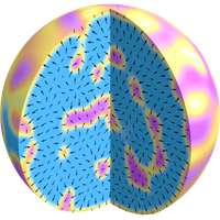
Chromatin, the functional form of DNA in cells, is a polymer immersed in the nucleoplasmic fluid inside the cell nucleus. It typically segregates into two phases: heterochromatin and euchromatin. Their segregation is often spatially patterned, with euchromatin enriched in the nuclear interior and heterochromatin concentrated near the nuclear envelope. We develop a multiphase continuum model that shows how mechanical forces and flows can drive chromatin phase segregation. Crosslinking proteins within heterochromatin generate contractile forces that pull it together into dropletlike domains that grow and merge over time. Confinement then favors their relocation and spreading along the nuclear envelope, producing a peripheral heterochromatin layer. Transcription-driven active forces in euchromatin drive nucleus-scale flows that deform and reorganize heterochromatin into many different morphologies. Overall, we present a theoretical framework linking active processes in chromatin to its mechanics, nucleus-wide patterning, and motion.
Article Text
Supplemental Material
References (111)
- B. Alberts, A. Johnson, J. Lewis, M. Raff, K. Roberts, and P. Walter, Molecular Biology of the Cell, 5th ed. (Garland Science, New York, 2008).
- T. Cremer and C. Cremer, Chromosome territories, nuclear architecture and gene regulation in mammalian cells, Nat. Rev. Genet. 2, 292 (2001).
- T. Misteli, Beyond the sequence: Cellular organization of genome function, Cell 128, 787 (2007).
- W. Bickmore and B. van Steensel, Genome architecture: Domain organization of interphase chromosomes, Cell 152, 1270 (2013).
- J. Gibcus and J. Dekker, The hierarchy of the 3D genome, Mol. Cell 49, 773 (2013).
- A. Pombo and N. Dillon, Three-dimensional genome architecture: Players and mechanisms, Nat. Rev. Mol. Cell Biol. 16, 245 (2015).
- L. Mirny and J. Dekker, Mechanisms of chromosome folding and nuclear organization: Their interplay and open questions, Cold Spring Harbor Perspect. Biol. 14, a040147 (2022).
- B. Bonev and G. Cavalli, Organization and function of the 3D genome, Nat. Rev. Genet. 17, 661 (2016).
- B. van Steensel and A. S. Belmont, Lamina-associated domains: Links with chromosome architecture, heterochromatin, and gene repression, Cell 169, 780 (2017).
- I. Solovei, K. Thanisch, and Y. Feodorova, How to rule the nucleus: Divide et impera, Curr. Opin. Cell Biol. 40, 47 (2016).
- A. Bizhanova and P. D. Kaufman, Close to the edge: Heterochromatin at the nucleolar and nuclear peripheries, Biochim. Biophys. Acta (BBA) 1864, 194666 (2021).
- D. Amiad-Pavlov, D. Lorber, G. Bajpai, A. Reuveny, F. Roncato, R. Alon, S. Safran, and T. Volk, Live imaging of chromatin distribution reveals novel principles of nuclear architecture and chromatin compartmentalization, Sci. Adv. 7, eabf6251 (2021).
- Y. Feodorova, M. Falk, L. A. Mirny, and I. Solovei, Viewing nuclear architecture through the eyes of nocturnal mammals, Trends Cell Biol. 30, 276 (2020).
- D. González-Acosta and M. Lopes, DNA replication and replication stress response in the context of nuclear architecture, Chromosoma 133, 57 (2024).
- J. Xu, H. Ma, J. Jin, S. Uttam, R. Fu, Y. Huang, and Y. Liu, Super-resolution imaging of higher-order chromatin structures at different epigenomic states in single mammalian cells, Cell Rep. 24, 873 (2018).
- C. D. Allis and T. Jenuwein, The molecular hallmarks of epigenetic control, Nat. Rev. Genet. 17, 487 (2016).
- C. Maison and G. Almouzni, HP1 and the dynamics of heterochromatin maintenance, Nat. Rev. Mol. Cell Biol. 5, 296 (2004).
- R. C. Allshire and H. D. Madhani, Ten principles of heterochromatin formation and function, Nat. Rev. Mol. Cell Biol. 19, 229 (2018).
- B. Mutlu, H.-M. Chen, J. J. Moresco, B. D. Orelo, B. Yang, J. M. Gaspar, S. Keppler-Ross, J. R. Yates, D. H. Hall, E. M. Maine, and S. E. Mango, Regulated nuclear accumulation of a histone methyltransferase times the onset of heterochromatin formation in C. elegans embryos, Sci. Adv. 4, eaat6224 (2018).
- A. R. Strom, A. V. Emelyanov, M. Mir, D. V. Fyodorov, X. Darzacq, and G. H. Karpen, Phase separation drives heterochromatin domain formation, Nature (London) 547, 241 (2017).
- A. G. Larson, D. Elnatan, M. M. Keenen, M. J. Trnka, J. B. Johnston, A. L. Burlingame, D. A. Agard, S. Redding, and G. J. Narlikar, Liquid droplet formation by suggests a role for phase separation in heterochromatin, Nature (London) 547, 236 (2017).
- A. G. Larson and G. J. Narlikar, The role of phase separation in heterochromatin formation, function, and regulation, Biochemistry 57, 2540 (2018).
- M. R. Hübner and D. L. Spector, Chromatin dynamics, Annu. Rev. Biophys. 39, 471 (2010).
- J. Lammerding, Mechanics of the nucleus, in Comprehensive Physiology (Wiley, New York, 2011), pp. 783–807.
- A. Zidovska, The rich inner life of the cell nucleus: Dynamic organization, active flows, and emergent rheology, Biophys. Rev. Lett. 12, 1093 (2020).
- I. Eshghi, J. A. Eaton, and A. Zidovska, Interphase chromatin undergoes a local sol-gel transition upon cell differentiation, Phys. Rev. Lett. 126, 228101 (2021).
- A. Janssen, S. U. Colmenares, and G. H. Karpen, Heterochromatin: Guardian of the genome, Annu. Rev. Cell Dev. Biol. 34, 265 (2018).
- E. M. Hildebrand and J. Dekker, Mechanisms and functions of chromosome compartmentalization, Trends Biochem. Sci. 45, 385 (2020).
- T. Misteli, The self-organizing genome: Principles of genome architecture and function, Cell 183, 28 (2020).
- A. Zidovska, D. A. Weitz, and T. J. Mitchison, Micron-scale coherence in interphase chromatin dynamics, Proc. Natl. Acad. Sci. U.S.A. 110, 15555 (2013).
- F.-Y. Chu, A. S. Clavijo, S. Lee, and A. Zidovska, Transcription-dependent mobility of single genes and genome-wide motions in live human cells, Nat. Commun. 15, 8879 (2024).
- A. Zidovska, The self-stirred genome: Large-scale chromatin dynamics, its biophysical origins and implications, Curr. Opin. Genet. Dev. 61, 83 (2020).
- R. Bruinsma, A. Y. Grosberg, Y. Rabin, and A. Zidovska, Chromatin hydrodynamics, Biophys. J. 106, 1871 (2014).
- I. Eshghi, A. Zidovska, and A. Y. Grosberg, Symmetry-based classification of forces driving chromatin dynamics, Soft Matter 18, 8134 (2022).
- I. Eshghi, A. Zidovska, and A. Y. Grosberg, Activity-driven phase transition causes coherent flows of chromatin, Phys. Rev. Lett. 131, 048401 (2023).
- I. Eshghi, A. Zidovska, and A. Y. Grosberg, Model chromatin flows: Numerical analysis of linear and nonlinear hydrodynamics inside a sphere, Eur. Phys. J. E 46, 69 (2023).
- M. Falk, Y. Feodorova, N. Naumova, M. Imakaev, B. R. Lajoie, H. Leonhardt, B. Joffe, J. Dekker, G. Fudenberg, I. Solovei, and L. A. Mirny, Heterochromatin drives compartmentalization of inverted and conventional nuclei, Nature (London) 570, 395 (2019).
- G. Bajpai, D. A. Pavlov, D. Lorber, T. Volk, and S. Safran, Mesoscale phase separation of chromatin in the nucleus, eLife 10, e63976 (2021).
- O. Adame-Arana, G. Bajpai, D. Lorber, T. Volk, and S. Safran, Regulation of chromatin microphase separation by binding of protein complexes, eLife 12, e82983 (2023).
- R. Laghmach, M. Di Pierro, and D. A. Potoyan, Four-dimensional mesoscale liquid model of nucleus resolves chromatin’s radial organization, PRX Life 2, 013006 (2024).
- N. Ganai, S. Sengupta, and G. I. Menon, Chromosome positioning from activity-based segregation, Nucleic Acids Res. 42, 4145 (2014).
- D. Michieletto, E. Orlandini, and D. Marenduzzo, Polymer model with epigenetic recoloring reveals a pathway for the de novo establishment and 3D organization of chromatin domains, Phys. Rev. X 6, 041047 (2016).
- J. Smrek and K. Kremer, Small activity differences drive phase separation in active-passive polymer mixtures, Phys. Rev. Lett. 118, 098002 (2017).
- C. Hult, D. Adalsteinsson, P. A. Vasquez, J. Lawrimore, M. Bennett, A. York, D. Cook, E. Yeh, M. G. Forest, and K. Bloom, Enrichment of dynamic chromosomal crosslinks drive phase separation of the nucleolus, Nucleic Acids Res. 45, 11159 (2017).
- Q. MacPherson, B. Beltran, and A. J. Spakowitz, Bottom–up modeling of chromatin segregation due to epigenetic modifications, Proc. Natl. Acad. Sci. U.S.A. 115, 12739 (2018).
- G. Shi, L. Liu, C. Hyeon, and D. Thirumalai, Interphase human chromosome exhibits out of equilibrium glassy dynamics, Nat. Commun. 9, 3161 (2018).
- M. D. Pierro, D. A. Potoyan, P. G. Wolynes, and J. N. Onuchic, Anomalous diffusion, spatial coherence, and viscoelasticity from the energy landscape of human chromosomes, Proc. Natl. Acad. Sci. U.S.A. 115, 7753 (2018).
- Q. MacPherson, B. Beltran, and A. J. Spakowitz, Chromatin compaction leads to a preference for peripheral heterochromatin, Biophys. J. 118, 1479 (2020).
- M. Di Stefano, H.-W. Nützmann, M. Marti-Renom, and D. Jost, Polymer modelling unveils the roles of heterochromatin and nucleolar organizing regions in shaping 3D genome organization in Arabidopsis thaliana, Nucleic Acids Res. 49, 1840 (2021).
- M. Girard, M. O. de la Cruz, J. F. Marko, and A. Erbaş, Heterogeneous flexibility can contribute to chromatin segregation in the cell nucleus, Phys. Rev. E 110, 014403 (2024).
- D. Saintillan, M. J. Shelley, and A. Zidovska, Extensile motor activity drives coherent motions in a model of interphase chromatin, Proc. Natl. Acad. Sci. U.S.A. 115, 11442 (2018).
- A. Mahajan, W. Yan, A. Zidovska, D. Saintillan, and M. J. Shelley, Euchromatin activity enhances segregation and compaction of heterochromatin in the cell nucleus, Phys. Rev. X 12, 041033 (2022).
- R. Das, T. Sakaue, G. Shivashankar, J. Prost, and T. Hiraiwa, How enzymatic activity is involved in chromatin organization, eLife 11, e79901 (2022).
- A. Goychuk, D. Kannan, A. K. Chakraborty, and M. Kardar, Polymer folding through active processes recreates features of genome organization, Proc. Natl. Acad. Sci. U.S.A. 120, e2221726120 (2023).
- S. Weady, D. B. Stein, A. Zidovska, and M. J. Shelley, Conformations, correlations, and instabilities of a flexible fiber in an active fluid, Phys. Rev. Fluids 9, 013102 (2024).
- V. I. P. Keizer, S. Grosse-Holz, M. Woringer, L. Zambon, K. Aizel, M. Bongaerts, F. Delille, L. Kolar-Znika, V. F. Scolari, S. Hoffmann, E. J. Banigan, L. A. Mirny, M. Dahan, D. Fachinetti, and A. Coulon, Live-cell micromanipulation of a genomic locus reveals interphase chromatin mechanics, Science 377, 489 (2022).
- R. J. Bearman and J. G. Kirkwood, Statistical mechanics of transport processes. XI. Equations of transport in multicomponent systems, J. Chem. Phys. 28, 136 (1958).
- F. M. Snell, R. Aranow, and R. A. Spangler, Statistical-mechanical derivation of the partial molecular stress tensors in isothermal multicomponent systems, J. Chem. Phys. 47, 4959 (1967).
- M. Doi and A. Onuki, Dynamic coupling between stress and composition in polymer solutions and blends, J. Phys. II (France) 2, 1631 (1992).
- M. Doi, Soft Matter Physics (Oxford University Press, New York, 2013).
- P. J. Foster, S. Fürthauer, M. J. Shelley, and D. J. Needleman, Active contraction of microtubule networks, eLife 4, e10837 (2015).
- J. Toner, Y. Tu, and S. Ramaswamy, Hydrodynamics and phases of flocks, Ann. Phys. (Amsterdam) 318, 170 (2005).
- M. Lachner, D. O’Carroll, S. Rea, K. Mechtler, and T. Jenuwein, Methylation of histone H3 lysine 9 creates a binding site for HP1 proteins, Nature (London) 410, 116 (2001).
- J. Padeken, S. P. Methot, and S. M. Gasser, Establishment of H3K9-methylated heterochromatin and its functions in tissue differentiation and maintenance, Nat. Rev. Mol. Cell Biol. 23, 623 (2022).
- C. R. Clapier, J. Iwasa, B. R. Cairns, and C. L. Peterson, Mechanisms of action and regulation of ATP-dependent chromatin-remodelling complexes, Nat. Rev. Mol. Cell Biol. 18, 407 (2017).
Here, the solvent pressure acts as a Lagrange multiplier that enforces the incompressibility condition in Eq. (11).
- D. Saintillan and M. J. Shelley, Instabilities, pattern formation, and mixing in active suspensions, Phys. Fluids 20, 123304 (2008).
- D. Saintillan and M. J. Shelley, Active suspensions and their nonlinear models, C.R. Phys. 14, 497 (2013).
- T. Gao, M. D. Betterton, A.-S. Jhang, and M. J. Shelley, Analytical structure, dynamics, and coarse graining of a kinetic model of an active fluid, Phys. Rev. Fluids 2, 093302 (2017).
- M. Doi and S. Edwards, The Theory of Polymer Dynamics (Oxford University Press, New York, 1986).
- S. Maddu, S. Weady, and M. J. Shelley, Learning fast, accurate, and stable closures of a kinetic theory of an active fluid, J. Comput. Phys. 504, 112869 (2024).
- R. Milo and R. Phillips, Cell Biology by the Numbers (Garland Science, New York, 2015).
- H. A. Shaban, R. Barth, L. Recoules, and K. Bystricky, Hi-D: Nanoscale mapping of nuclear dynamics in single living cells, Genome Biol. 21, 95 (2020).
- R. Barth, K. Bystricky, and H. A. Shaban, Coupling chromatin structure and dynamics by live super-resolution imaging, Sci. Adv. 6, eaaz2196 (2020).
- T. A. Daugird, Y. Shi, K. L. Holland, H. Rostamian, Z. Liu, L. D. Lavis, J. Rodriguez, B. D. Strahl, and W. R. Legant, Correlative single molecule lattice light sheet imaging reveals the dynamic relationship between nucleosomes and the local chromatin environment, Nat. Commun. 15, 4178 (2024).
- J. P. Siebrasse, R. Veith, A. Dobay, H. Leonhardt, B. Daneholt, and U. Kubitscheck, Discontinuous movement of mRNP particles in nucleoplasmic regions devoid of chromatin, Proc. Natl. Acad. Sci. U.S.A. 105, 20291 (2008).
- L. Liang, X. Wang, D. Xing, T. Chen, and W. R. Chen, Noninvasive determination of cell nucleoplasmic viscosity by fluorescence correlation spectroscopy, J. Biomed. Opt. 14, 024013 (2009).
- F. Erdel, M. Baum, and K. Rippe, The viscoelastic properties of chromatin and the nucleoplasm revealed by scale-dependent protein mobility, J. Phys. Condens. Matter 27, 064115 (2015).
- M. Hertzog and F. Erdel, The material properties of the cell nucleus: A matter of scale, Cells 12, 1958 (2023).
- K. J. Burns, G. M. Vasil, J. S. Oishi, D. Lecoanet, and B. P. Brown, Dedalus: A flexible framework for numerical simulations with spectral methods, Phys. Rev. Res. 2, 023068 (2020).
- See Supplemental Material at http://link.aps.org/supplemental/10.1103/8n8h-7gx6 for additional figures and videos.
- R. A. Simha and S. Ramaswamy, Hydrodynamic fluctuations and instabilities in ordered suspensions of self-propelled particles, Phys. Rev. Lett. 89, 058101 (2002).
- M. C. Marchetti, J.-F. Joanny, S. Ramaswamy, T. B. Liverpool, J. Prost, M. Rao, and R. A. Simha, Hydrodynamics of soft active matter, Rev. Mod. Phys. 85, 1143 (2013).
- I. Solovei, A. Wang, K. Thanisch, C. Schmidt, S. Krebs, M. Zwerger, T. Cohen, D. Devys, R. Foisner, L. Peichl, H. Herrmann, H. Blum, D. Engelkamp, C. Stewart, H. Leonhardt, and B. Joffe, LBR and lamin A/C sequentially tether peripheral heterochromatin and inversely regulate differentiation, Cell 152, 584 (2013).
- J. Kind, L. Pagie, H. Ortabozkoyun, S. Boyle, S. de Vries, H. Janssen, M. Amendola, L. Nolen, W. Bickmore, and B. van Steensel, Single-cell dynamics of genome-nuclear lamina interactions, Cell 153, 178 (2013).
- J. Miné-Hattab and I. Chiolo, Complex chromatin motions for DNA repair, Front. Genet. 11, 800 (2020).
- P. M. Krawczyk, T. Borovski, J. Stap, A. Cijsouw, R. ten Cate, J. P. Medema, R. Kanaar, N. A. P. Franken, and J. A. Aten, Chromatin mobility is increased at sites of DNA double-strand breaks, J. Cell Sci. 125, 2127 (2012).
- H. Lans, J. H. J. Hoeijmakers, W. Vermeulen, and J. A. Marteijn, The DNA damage response to transcription stress, Nat. Rev. Mol. Cell Biol. 20, 766 (2019).
- J. A. Eaton and A. Zidovska, Structural and dynamical signatures of local DNA damage in live cells, Biophys. J. 118, 2168 (2020).
- F. G. Fernández, J. Park, C. Chapuis, E. P. Jurado, V. Imburchia, R. Smith, E. J. Longarini, A. Taddei, C. Hubert, N. Sokolovska, I. Matić, S. Huet, and J. Miné-Hattab, Single nucleosome imaging reveals principles of transient multiscale chromatin reorganization triggered by histone ADP-ribosylation at DNA lesions, Nat. Commun. 16, 6652 (2025).
- K. Maeshima, T. Matsuda, Y. Shindo, H. Imamura, S. Tamura, R. Imai, S. Kawakami, R. Nagashima, T. Soga, H. Noji, K. Oka, and T. Nagai, A transient rise in free ions released from ATP-Mg hydrolysis contributes to mitotic chromosome condensation, Curr. Biol. 28, 444 (2018).
- R. H. Wright, F. L. Dily, and M. Beato, ATP, , nuclear phase separation, and genome accessibility, Trends Biochem. Sci. 44, 565 (2019).
- K. Maeshima, S. Iida, and S. Tamura, Physical nature of chromatin in the nucleus, Cold Spring Harbor Perspect. Biol. 13, a040675 (2021).
- A. Zidovska, Chromatin: Liquid or solid?, Cell 183, 1737 (2020).
- H. Strickfaden, T. O. Tolsma, A. Sharma, D. A. Underhill, J. C. Hansen, and M. J. Hendzel, Condensed chromatin behaves like a solid on the mesoscale in vitro and in living cells, Cell 183, 1772 (2020).
- https://github.com/sarautu/nucleoplasm.
- D. L. Levy and R. Heald, Nuclear size is regulated by Importin and NTF2 in Xenopus, Cell 143, 288 (2010).
- S. Machida, Y. Takizawa, M. Ishimaru, Y. Sugita, S. Sekine, J. ichi Nakayama, M. Wolf, and H. Kurumizaka, Structural basis of heterochromatin formation by human HP1, Mol. Cell 69, 385 (2018).
- M. M. Keenen, D. Brown, L. D. Brennan, R. Renger, H. Khoo, C. R. Carlson, B. Huang, S. W. Grill, G. J. Narlikar, and S. Redding, HP1 proteins compact DNA into mechanically and positionally stable phase separated domains, eLife 10, e64563 (2021).
- D. N. Itzhak, S. Tyanova, J. Cox, and G. H. Borner, Global, quantitative and dynamic mapping of protein subcellular localization, eLife 5, e16950 (2016).
- K. P. Müller, F. Erdel, M. Caudron-Herger, C. Marth, B. D. Fodor, M. Richter, M. Scaranaro, J. Beaudouin, M. Wachsmuth, and K. Rippe, Multiscale analysis of dynamics and interactions of heterochromatin protein 1 by fluorescence fluctuation microscopy, Biophys. J. 97, 2876 (2009).
- L. Schmiedeberg, K. Weisshart, S. Diekmann, G. M. zu Hoerste, and P. Hemmerich, High- and low-mobility populations of HP1 in heterochromatin of mammalian cells, Mol. Biol. Cell 15, 2819 (2004).
- Y. Tseng, J. S. H. Lee, T. P. Kole, I. Jiang, and D. Wirtz, Micro-organization and visco-elasticity of the interphase nucleus revealed by particle nanotracking, J. Cell Sci. 117, 2159 (2004).
- S. M. Görisch, M. Wachsmuth, C. Ittrich, C. P. Bacher, K. Rippe, and P. Lichter, Nuclear body movement is determined by chromatin accessibility and dynamics, Proc. Natl. Acad. Sci. U.S.A. 101, 13221 (2004).
- F. M. Hameed, M. Rao, and G. V. Shivashankar, Dynamics of passive and active particles in the cell nucleus, PLoS One 7, e45843 (2012).
- H. D. Ou, S. Phan, T. J. Deerinck, A. Thor, M. H. Ellisman, and C. C. O’Shea, ChromEMT: Visualizing 3D chromatin structure and compaction in interphase and mitotic cells, Science 357, eaag0025 (2017).
- E. Miron, R. Oldenkamp, J. M. Brown, D. M. S. Pinto, C. S. Xu, A. R. Faria, H. A. Shaban, J. D. P. Rhodes, C. Innocent, S. de Ornellas, H. F. Hess, V. Buckle, and L. Schermelleh, Chromatin arranges in chains of mesoscale domains with nanoscale functional topography independent of cohesin, Sci. Adv. 6, eaba8811 (2020).
- C. Bustamante, W. Cheng, and Y. X. Mejia, Revisiting the central dogma one molecule at a time, Cell 144, 480 (2011).
- G. J. Wuite, S. B. Smith, M. Young, D. Keller, and C. Bustamante, Single-molecule studies of the effect of template tension on T7 DNA polymerase activity, Nature (London) 404, 103 (2000).
- E. A. Galburt, S. W. Grill, A. Wiedmann, L. Lubkowska, J. Choy, E. Nogales, M. Kashlev, and C. Bustamante, Backtracking determines the force sensitivity of RNAP II in a factor-dependent manner, Nature (London) 446, 820 (2007).
- E. A. Abbondanzieri, W. J. Greenleaf, J. W. Shaevitz, R. Landick, and S. M. Block, Direct observation of base-pair stepping by RNA polymerase, Nature (London) 438, 460 (2005).

